Embryo development and evaluation, part 1

An important part of being an embryologist is being able to evaluate what an embryo looks like. We do this because embryos that “look better” have a better chance of working compared to embryos that don’t look as good.
This is the 1st part of a 3 part series where I’ll talk about the different factors that go into this evaluation.
In this segment I’ll talk about embryo development before I get into the grading of cleavage stage embryos and then finally the grading of blastocysts.
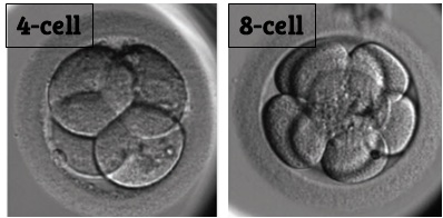
An embryo begins when a sperm fertilizes an egg cell (aka an oocyte - pronounced “oh-cyte” or “oh-oh-cyte”). This is sometimes called the “pronuclear stage”. Once the egg is fertilized, this single cell divides (or cleaves) after about 24 hours to make two cells. From 2 cells, each cell then divides again to form 4 cells (Day 2), and then again to make 8 cells (Day 3).
This stage is sometimes called the “cleavage stage” because the cells of the embryo cleave to make more cells. You can see an example of a 4 cell and 8 cell embryo below during the cleavage stage.

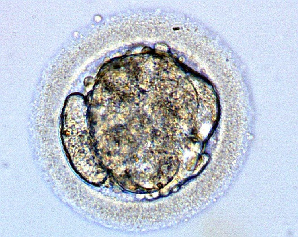
After Day 3, the embryo typically will start to form a morula. This is a specialized structure that reminds me of a cocoon and usually forms around Day 4 after insemination. All the cells that were formed during the cleavage stage start to come together to form a mass of cells.
Here’s an image of a morula below. Notice how it’s hard to even tell there’s individual cells anymore (with the exception of the cell on the left)!

I liken the morula to a cocoon because on the inside, something very special is happening!
After the morula forms, the embryo develops into the next stage called a “blastocyst” or “blast” for short. This usually happens after about 5-7 days from insemination of the egg.
A blastocyst is named because it contains a blastocoel (pronounced blasto-seal). This is a sac of water inside the embryo that can help the embryo get bigger so it can hatch from its zona (the shell surrounding the embryo).
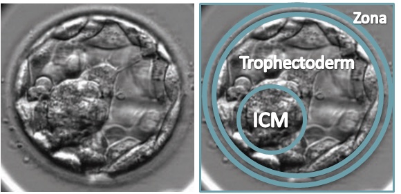
Blastocysts have two special features that we can use to evaluate their quality. The inner cell mass (or ICM) is a mass of cells that eventually forms the fetus. The trophectoderm are the cells all around the ICM that eventually form the placenta.
Here’s an image of a blastocyst below with these features indicated:

As the embryo develops further, it eventually hatches from its shell (zona). Now the cells of the embryo can directly interact with the uterine lining to implant and hopefully form a pregnancy.
Below is an overview of the different stages of an embryo’s development along with the day of the embryo’s development after insemination:

That’s it for this segment! Join me next time and I’ll get into how we can grade cleavage stage embryos to determine which embryo has the highest potential.
Can’t wait? I offer evaluations and a video course to help people better understand their embryo pictures - check it out here: https://www.remembryo.com/embryo-grading-request/
Also check me out on Instagram! https://www.instagram.com/embryomanofficial/
Recent blog posts
Excerpt from the memoir “How to Get a Girl Pregnant”
Waiting. Waiting. Waiting. Can I fill up the day with 16 separate tasks so that I don’t think about waiting?
Just Adopt
There’s really no escaping it. When you’re facing infertility, other people getting into your business begins to feel pretty routine.
Four things I’ve learned about having a baby in my 40s
As many of us that have struggled to get pregnant know, life doesn’t always go the way you expect it to.


